

Eye Anatomy And Diagram

Understanding your eyes

Here's a handy guide to the anatomy of your eye, so you can know what eye experts are talking about.
Your eyes are one of the most intricate systems in your body. Their inner workings are miraculous, but don't be intimidated. The independent vision specialists at or next to your local Pearle Vision EyeCare Centre are always ready to explain things in everyday terms.
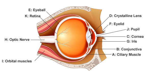
- A: Ciliary Muscle Creates focus by adjusting the shape of your eye. Over time loses its elasticity (which is why we all eventually need reading glasses).
- B: Conjunctiva A clear membrane covering the front of your eye and inner eyelids. Keeps eyes moist and is first line of defense against infection.
- C: Cornea Covers the front of the eye. It is the first and most powerful lens in the eye's optical system. Needs to be protected from accidents, infections or genetic defects.
- D: Crystalline Lens Just behind the cornea: it keeps the world in focus on your retina. A cataract is the clouding of this lens.
- E: Eyeball Measuring about 1 inch across in diameter. If yours is a little larger, you'll be nearsighted (myopic); if it's smaller, you're farsighted (hyperopic).
- F: Eyelid Protects and lubricates our eyes.
- G: Iris This is the colored part of your eye. This ring of muscle fibers contracts and expands, to open and close your pupil, based on surrounding light.
- H: Optic Nerve Connects your eye and brain with roughly 1.2 million nerve fibers.
- I: Orbital Muscles Six muscles are in charge of eye movement. Four control up, down, left and right. The other two control your eyes when your head tilts.
- J: Pupil The pupil is the hole in the center of the iris that light passes through. The iris controls its size.
- K: Retina Converts light into electrical signals and sends them to the brain through the optic nerve.